Impact of physicochemical properties of inorganic nanoparticles on their autophagic effects and toxicity (2014-2017)
Introduction
Many inorganic nanoparticles (INPs) have recently been synthesized and used in the biological research area. It has showed that they likely induce the the autophagic effects and thus lead to a dysfunction of our cellular system, due to their unique physicochemical properties. However, it is still unclear whether these properties (such as size, morphology, surface modification and dispersity) can significantly induce the autophagic effects.
Main investigations
In this project, Hualu, as a master graduate student supervised by Professor Jinhao Gao at Xiamen University, synthesized gold, iron oxide, and silica oxide nanoparticles with controllable properties, and generally found that inorganic nanoparticles could modulate autophagic effect by their unique properties, particularly including their dispersity, shape, and size [1].
- Shape
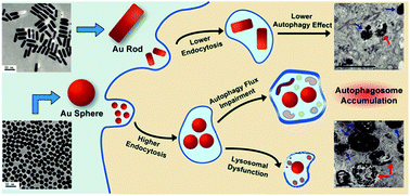
We synthesized the gold nanoparticles with different shapes, and compared their cellular interactions, uptakes, and autophagic effects. The results revealed that gold nanoparticles could modulate autophagy in a shape-dependent manner. Western blot assays and confocal images confirmed that nanospheres caused more autophagosome accumulation than nanorods, which were highly correlated with the difference in cellular uptakes. Please check out the publication [2] for more details.
- Size
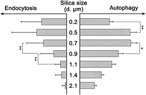
We also showed that silica sub-microspheres (0.1 to 2.1 μm in diameter) induced autophagy, which depended on the levels of cellular endocytosis. Due to the suitable size for cellular endocytosis, 0.5–0.7 μm silica particles induced the highest levels of autophagy among particles from 0.1 to 2.1 μm in diameter. Changes in cellular endocytosis of silica sub-microspheres lead to alteration of autophagy levels. Furthermore, dephosphorylation of FOXO3A and subsequent translocation to the nucleus could be associated with this autophagy process. Our results revealed that silica sub-microspheres induced autophagy, and emphasized the potential risk of endocytosis of fine particles or other non-degradable materials. Please check out the publication [3] for more details.
- Dispersity
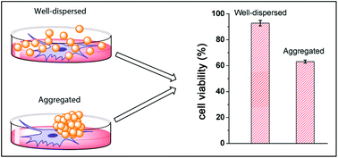
We selected gold nanoparticles as an example and investigated the role of dispersity in the autophagic effect and a potential cytotoxicity. Our results indicated that the cytotoxicity of aggregated gold nanoparticles is significantly higher than well-dispersed ones. The dispersity-dependent cytotoxicity could be related to the increase of cellular endocytosis and reactive oxygen species. Please check out the publications [4] and [5] for more details.
Reference
Zhou, H.‡; Gong, X.‡; Lin, H.‡; Chen, H.; Huang, D.; Li, D.; Shan, H.; Gao, J. Gold nanoparticles impair autophagy flux through shape-dependent endocytosis and lysosomal dysfunction. Journal of Materials Chemistry B 2018, 6 (48), 8127-8136. DOI: https://doi.org/10.1039/C8TB02390E.
Huang, D.‡; Zhou, H.‡; Liu, H.; Gao, J. The cytotoxicity of gold nanoparticles is dispersity-dependent. Dalton Transactions 2015, 44 (41), 17911-17915. DOI: https://doi.org/10.1039/C5DT02118A.
Huang, D.; Zhou, H.; Gao, J. Nanoparticles modulate autophagic effect in a dispersity-dependent manner. Scientific Reports 2015, 5 (1), 14361. DOI: https://doi.org/10.1038/srep14361.
Huang, D.; Zhou, H.; Gong, X.; Gao, J. Silica sub-microspheres induce autophagy in an endocytosis dependent manner. RSC Advances 2017, 7 (21), 12496-12502. DOI: https://doi.org/10.1039/C6RA26649E.


